
blog tambre
Mosaic Embryos: What do you know?
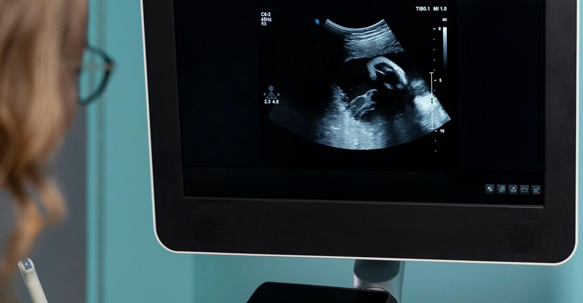
Table of contents
Those of you who are familiar with this blog will know that we use this this space to cover topics that you might possibly come across at some point in your fertility treatment. Today is the turn of mosaic embryos, has anyone ever heard of them?
What is a mosaic embryo?
karyotype refers to an individual’s complete set of chromosomes. We all have 46 chromosomes (23 pairs) of which half are inherited from your mother and half from your father. In order for an embryo to develop properly, it needs to contain this pairing of chromosomes.
A mosaic embryo is one that contains 2 or more cells with different genetic content, that is, it has a mix of normal and abnormal chromosomes. Mosaic embryos can be classified according to the number of chromosomes involved (simple or complex), type of aneuploidism, that is, whether it affects the entire chromosome (complete), a segment (partial) or degree of mosaicism (moderate or extended).
How do we identify mosaic embryos?
Mosaicism is detected through a technique called Pre -implantation Genetic Diagnosis, which analyzes the genetic content of cells biopsied in the trofoectoderm (outer layer of the embryo). This analysis is performed using Next Generation Sequencing (NGS) techniques.
Mosaicism appears as the result of an error in chromosomic segregation that could be have occurred in the sperm, ovary or by errors during the time the embryo divides into two.
It is generally accepted in most studies that the morphology of the embryo is not related to its genetic content – that is tos ay, that the microscopic appearance of an embryo is only mildly related to its viability. This is the reason why we at Tambre employ the Pre -implantation Genetic Diagnosis technique.
What does science say about mosaic embryos?
There has been a lot of research into the area of mosaic embryos. Thanks to various scientific interventions, extensive guidelines for the management of mosaic embryos have been developed. In 2015, the first baby was delivered after the transfer of a mosaic embryo and since then, more cases have been managed.
It is always advisable to seek appropriate genetic advice from a geneticist who can explain and clarify all the risks that could be associated with chromosomal alteration. The transfer of these embryos will always take place when there are no euploid embryos to transfer.
The possibility of being able to transfer a mosaic embryo or not depends, accordingly on the view of the geneticist’s and their opinion of the type of mosaicism that an embryo has. Directors of the International Society for Pre -implanted Genetic Diagnosis (PGDIS) suggest that priority should be given to the transfer of embryos based on the levels of mosaicism as well as the number and specific chromosomes involved. Mosaic embryos with affected chromosomes 13, 15, 16, 18, 21, 22, X and Y are considered “high risk” given their possible connection with uniparental isodisomy (both chromosomes or chromosomal regions are identical), a severe restriction of intrauterine growth and the possibility of a aneuploide birth (where there is the presence of an abnormal number of chromosomes in a cell).
When should a mosaic embryo be transferred and what procedure should be followed
Transfer of mosaic-only embryos is acceptable if there are no embryos considered euploid after receiving DGP (or PGT-A) or if the number of euploid embryos has increased. However, it should be noted that there is a type of mosaic embryo called “aneuploid mosaic” and that should not be transferred in any situation. These embryos are those that contain two or more different abnormal cell lines. and therefore are not viable.
When carrying out the transfer of a mosaic embryo, the instructions to follow before and after the intervention are exactly the same as if the embryo was euploid. Whenever there is a positive beta, specialists recommend the implementation of a prenatal crib technique during the first trimester to determine if the embryo has developed in an aneuploidal sense.
This test would form part of what is called an invasive prenatal crib and is called a corial biopsy. This test consists of taking a sample of placenta (coral velocities) and it can be performed vaginally (transcervical) or transabdominal. It can be done between 10 and 14+5 weeks of treatment, because it offers the possibility of getting a diagnosis earlier than if an amniocentesis is performed.